By Brenna Mackay
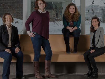
Carleton University researchers in physics and engineering are joining forces with biologists and medical practitioners to change how diseases are diagnosed and treated.
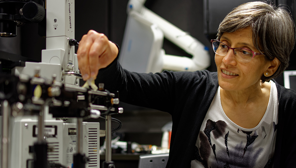
Physics Prof. Sangeeta Murugkar leads Carleton University’s Laboratory for Laser-Assisted Medical Physics and Engineering, which aims to develop optical imaging techniques for disease diagnosis and monitoring.
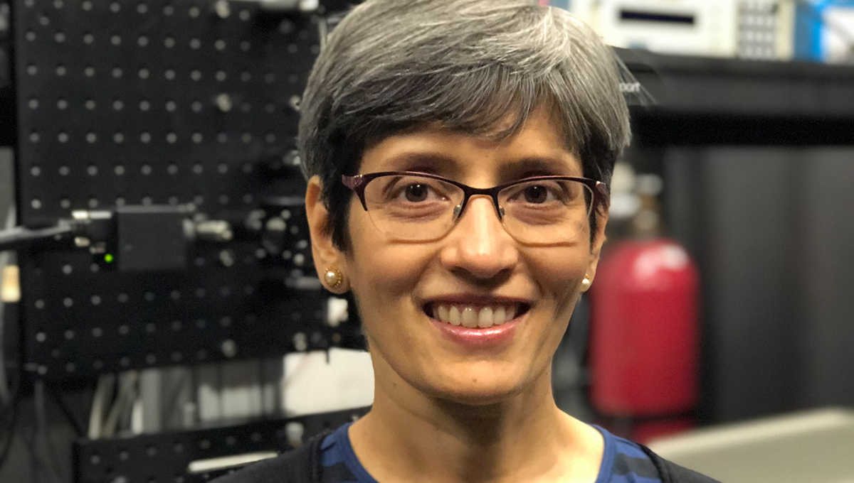
Prof. Sangeeta Murugkar
“There are two major goals,” explains Murugkar. “To develop rapid non-invasive, yet highly accurate medical diagnostics, as well as to enable fundamental biological discovery.”
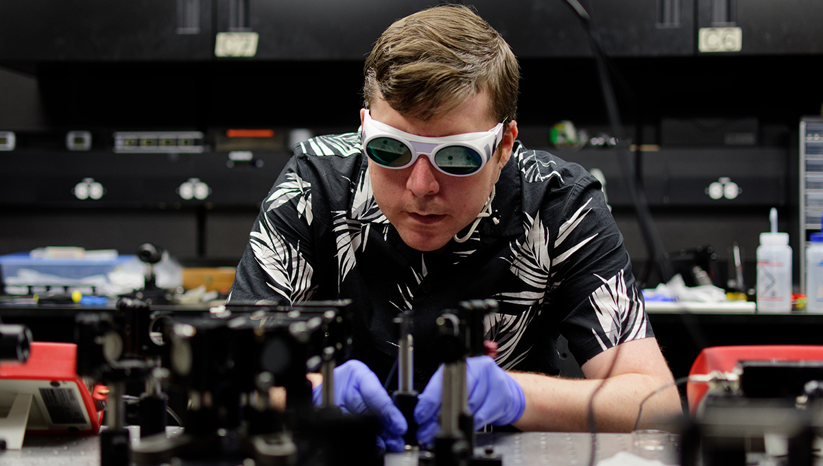
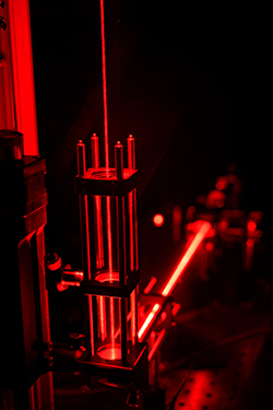
Working towards this, the lab uses a special optical effect called the Raman effect.
“When light interacts with matter, the colour or wavelength of most of the light does not change,” she says.
However, the wavelength of a very small fraction of the light changes when the incident light beam excites vibrations of molecules, due to the Raman effect. By analyzing these small colour changes or shifts in energy, her team can trace them back to chemical bonds or molecules in the sample.
“It’s kind of like chemical fingerprinting,” Murugkar adds. “Imagine if a doctor could shine light at the patient’s body and immediately diagnose the disease. This is rapid non-invasive optical biopsy.”

Prof. Leila Mostaço-Guidolin
Prof. Leila Mostaço-Guidolin is a new researcher at Carleton who joined the Systems and Computer Engineering Department in January 2020. She oversees the Carleton University (bio)Printing & Imaging (CUPIM) lab , an interdisciplinary research group developing novel 3D tissue models by using 3D bioprinting technology and optical imaging to monitor and characterize biochemical and structural aspects of biological tissue in non-invasive ways.
“It works in a comparable way to the regular 3D printing that everyone is talking about,” says Mostaço-Guidolin.
“But instead of using plastic or just synthetic materials, we can add proteins and cells to mimic the composition of the tissues we find in the body.”
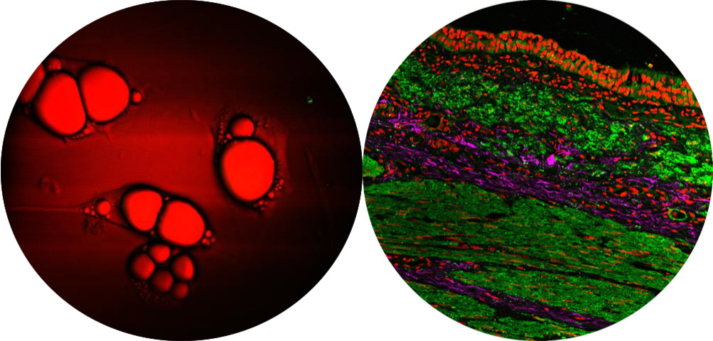
The Extracellular Matrix
By 3D bioprinting sample models, her team can make them as simple or as complicated as they want. This allows them to have full control over the structures and quickly analyze them.
 She explains that the human body is made up of many cells that must attach to a structure to form things like tissues and organs. The network that the cells use to form structure is called the extracellular matrix, which is made up of different protein fibres and other components.
Her work seeks to understand what changes occur when tissue is exposed to different chemical and physical stimuli.
“Once we have a better understanding about the physical and chemical properties of the matrix, how the cells are attached to it, we can then design new types of drugs to hopefully address these changes,” Mostaço-Guidolin says.
“Using certain optical imaging technologies, we can look at these structures without having to add stains or other chemicals, so we can really get a very nice picture of the samples, without disrupting the micro-environment the cells are in.”
Mostaço-Guidolin and Murugkar agree that their research would not be possible without collaboration across fields.
“More than ever, scientific research has become very multidisciplinary in all aspects,” says Mostaço-Guidolin. “It’s important to work together to explore current ideas and ways to integrate knowledge that you wouldn’t otherwise have.”
To learn more about Life Science research at Carleton, visit the website.
Tuesday, April 27, 2021 in Faculty of Science, Physics, Research
Share: Twitter, Facebook